
Fig. 4
- ID
- ZDB-FIG-111026-16
- Publication
- Lu et al., 2011 - Induction of apoptosis and inhibition of cell growth by tbx5 knockdown contribute to dysmorphogenesis in Zebrafish embryos
- Other Figures
- All Figure Page
- Back to All Figure Page
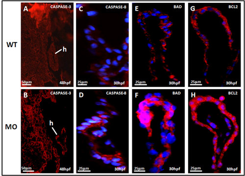
Sagittal view of the heart in tbx5 knockdown embryos by immunohistochemical studies of heart. A-B: Caspase-3 (red) expression could been slightly seen in wild-type (WT) embryos (A), but caspase-3 was more induced in knockdown embryos at 48 h post-fertilization (hpf) though the expression was not so strong (B). Caspase-8 (blue) was more induced in knockdown embryos (D) at 48 h post-fertilization (hpf) than the expression in wild-type (C). The proapoptotic factor, BAD (red), was enhanced in tbx5 deficiency embryos (F) with the nucleus stained by DAPI (blue); but there was almost no BAD expression in WT embryos (E). Compared to the WT (G), embryos with tbx5 deficiency showed a significant increase in BLC2 expression (H). The anterior of embryos is to the left. h, heart; MO, tbx5-MO-treated embryos. |
| Gene: | |
|---|---|
| Antibody: | |
| Fish: | |
| Knockdown Reagent: | |
| Anatomical Term: | |
| Stage: | Long-pec |