|
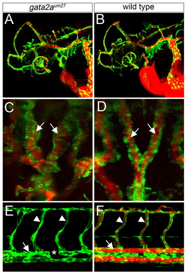
gata2aum27 mutant embryos display defects in aorta morphogenesis. Confocal microangiography using Quantum Dots in Tg(kdrl:egfp)la116 transgenic zebrafish embryos. Endothelial cells are green and vessel perfusion is red. (A,B) Cranial blood vessels in gata2aum27 mutant (A) and wild-type (B) embryos. Lateral views, anterior to the left, dorsal is up. (C,D) Lateral dorsal aortae (arrows) in gata2aum27 mutant (C) and wild-type (D) embryos. Dorsal views, anterior is up. (E,F) Trunk blood vessels in gata2aum27 mutant (E) and wild-type (F) embryos. Lateral views, anterior to the left, dorsal is up. Segmental vessels are indicated by arrowheads and the dorsal aorta by an arrow; the asterisk indicates a region of aorta that failed to form.
|