
Fig. S1
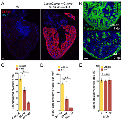
Robust, dose-dependent cardiomyocyte ablation. (A) Anti-DsRed staining to indicate mCherry expression in most if not all ventricular and atrial cardiomyocytes of transgenic animals (bactin2:loxp-mCherry-STOP-loxp-DTA). (B) Phalloidin staining indicates heavy cardiac myofiber loss at 7 days following a single 4-HT injection (bottom), compared with vehicle control (top). (C,D) Quantification of myofiber area by MHC (C) or Mef2 (D) staining in Z-CAT fish incubated with vehicle (n=5) or two different concentrations of 4-HT (20 nM, n=5; 500 nM, n=3) for 12 hours. Ablation was assessed at 7 days after incubation in 4-HT. Mean±s.e.m. **P<0.005, *P<0.05, Studentýs t-test. (E) Ventricular section surface area was measured using Openlab software, using the three largest sections from each ventricle. Five or six animals were assessed for each group. Scale bars: 50 μm. |