
Fig. 5
- ID
- ZDB-FIG-110909-25
- Publication
- Lewis et al., 2011 - Celsr3 Is Required for Normal Development of GABA Circuits in the Inner Retina
- Other Figures
- All Figure Page
- Back to All Figure Page
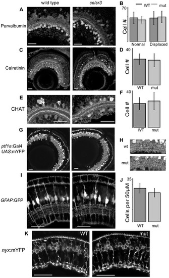
Cell localization and IPL organization are unchanged in the celsr3 mutant. A) α-parvalbumin labels all displaced amacrine cells and a subpopulation of amacrines in the INL. B) counts of parvalbumin cells, displaced and normal amacrines were counted separately C) α-calretinin labels a subpopulation of amacrines in the INL and all ganglion cells. D) Counts of calretinin positive amacrine cells. E) α-CHAT stains a subpopulation of amacrine cells that laminate in 2 major and 2 minor sublaminae within the IPL. All sublaminae are present in the mutant. F) counts of CHAT positive amacrine cells in the INL G) Tg(ptf1a:Gal4VP16, UAS:mYFP) animals express mYFP in all amacrine and horizontal cells. H) Close ups of the IPL in Tg(ptf1a:Gal4VP16, UAS:mYFP) animals I) Images of Tg(GFAP:GFP) animals, which express GFP in all Müller cells. J) Counts of Müller cells per 50 μM. K) Tg(nyx:mYFP) animals express mYFP in the ON-bipolar cells. Scale bars are 20 μm. |
| Genes: | |
|---|---|
| Fish: | |
| Anatomical Terms: | |
| Stage: | Day 6 |
| Fish: | |
|---|---|
| Observed In: | |
| Stage: | Day 6 |