
Fig. 3
- ID
- ZDB-FIG-110812-4
- Publication
- Paridaen et al., 2011 - The nucleolar GTP-binding proteins Gnl2 and nucleostemin are required for retinal neurogenesis in developing zebrafish
- Other Figures
- All Figure Page
- Back to All Figure Page
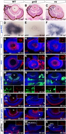
Delayed RGC differentiation in the gnl2 and ns mutant retina. (A–C) Transverse toluidine-blue stained sections of WT (A), gnl2 (B) and ns (C) showing incomplete formation of GCL, INL and ONL at 72 hpf (arrowheads). Arrow indicates optic nerve head. (D–F) Expression of ath5 marks retinal progenitors prior to cell cycle exit. At 36 hpf, ath5 is absent apart from a few cells in gnl2 mutants (E; n = 7 embryos), whereas its expression is reduced, but is normally patterned in ns mutants (F; n = 6). Dashed circle indicates the outline of the lens. (G–L) Confocal images of immunolabelings to the RGC marker zn8 (red) with DAPI nuclear counterstaining (blue) at 48 (G–I) and 72 (J–L) hpf. In gnl2 mutants, the GCL is disorganized and reduced in width at both 48 hpf (H; n = 11) and 72 hpf (K; n = 25). In ns mutants, formation of the GCL layer is incomplete at 48 hpf (I; n = 22), but has markedly improved at 72 hpf (L; n = 19). Insets show single zn8 fluorescence views. Dashed circles indicate the outline of the lens; the additional outer circle in (K) delineates the outline of the eye. (M–O) Single confocal planes of Hu/D (green) and zn8 (red) labeling with DAPI nuclear counterstaining (blue), which labels RGCs and differentiated amacrine cells, and RGCs only, respectively, in gnl2 (N; n = 5) and ns mutants (O; n = 6) at 48 hpf. Arrowhead shows amacrine cells that are labeled with HuC/D, but not with zn8. Dashed line shows boundary of GCL, dotted line marks boundary of amacrine cells in the INL. Insets show single HuC/D and zn8 fluorescence views. (P–R) At 96 hpf, differentiated photoreceptor cells marked by zpr1 staining in red with DAPI nuclear counterstaining (blue) are present in the central gnl2 mutant retina only (boundaries indicated with arrowheads in Q; n = 8). In ns mutants, photoreceptor cells have differentiated and are present throughout the outer nuclear layer (R; n = 5). Insets show single zpr1 fluorescence views. (S–U) At 96 hpf, Müller glial cells, marked by zrf1 staining in red with DAPI nuclear counterstaining (blue), have developed relatively normally in ns mutants (U; n = 11), but are disorganized and reduced in gnl2 mutants (arrowhead in T; n = 9). Insets show single zrf1 fluorescence views. (V–X) Differentiation of retinal cell types, such as RGCs (n = 5; V; compare to J), photoreceptor cells (n = 4; W, compare to P) and Müller glial cells (n = 3; X, compare to S) is severely affected in gnl2/ns compound mutants as compared to WTs. Inset show single fluorescence view. (A–C) Lateral up, anterior to the left. (D–L) Lateral view, anterior to the left and dorsal up. (M–X) Dorsal view, with anterior to the left. GCL, ganglion cell layer; INL, inner nuclear layer; le, lens; ONL, outer nuclear layer. Scalebar 25 μm. |
| Gene: | |
|---|---|
| Antibodies: | |
| Fish: | |
| Anatomical Terms: | |
| Stage Range: | Prim-25 to Day 4 |
| Fish: | |
|---|---|
| Observed In: | |
| Stage Range: | Prim-25 to Day 4 |
Reprinted from Developmental Biology, 355(2), Paridaen, J.T., Janson, E., Utami, K.H., Pereboom, T.C., Essers, P.B., van Rooijen, C., Zivkovic, D., and Macinnes, A.W., The nucleolar GTP-binding proteins Gnl2 and nucleostemin are required for retinal neurogenesis in developing zebrafish, 286-301, Copyright (2011) with permission from Elsevier. Full text @ Dev. Biol.