
Fig. 5
- ID
- ZDB-FIG-110706-19
- Publication
- Budi et al., 2011 - Post-embryonic nerve-associated precursors to adult pigment cells: genetic requirements and dynamics of morphogenesis and differentiation
- Other Figures
- All Figure Page
- Back to All Figure Page
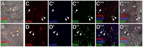
Proliferative extra-hypodermal cells revealed by post-embryonic EdU incorporation. All images from transverse sections of wild-type larvae staged as in Figure 3 (see Figure S4 for comparisons with erbb3b mutant larvae). (A,B) Merged images showing cells (arrowheads) within the myotomes labeled for either mitfa::GFP (red in A) or sox10::GFP (red in B) as well as EdU (green). (C,D) Cells within the lateral myotomes (m) and near the hypodermis (C) or at the base of the anal fin (f in D) labeled for mitfa::GFP (red), foxd3 (blue), and EdU (green). Merged views show fluorescence images or fluorescence images with brightfield overlays. Arrowheads, triple-labeled cells. m, myotome. e, epidermis. |