
Fig. 1
- ID
- ZDB-FIG-110608-34
- Publication
- Popgeorgiev et al., 2011 - The Apoptotic Regulator Nrz Controls Cytoskeletal Dynamics via the Regulation of Ca(2)+ Trafficking in the Zebrafish Blastula
- Other Figures
- All Figure Page
- Back to All Figure Page
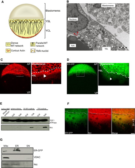
Nrz Protein Is Located into YSL Mitochondria and ER (A) Schematic drawing of a zebrafish embryo representing a simplified model of yolk organization. YSL contains a large number of nuclei present beneath the blastoderm. The region populated with yolk nuclei presents a dense network of microtubules from which originates a parallel microtubule network oriented along the animal-vegetal axis and extending into the yolk cell layer. MT, microtubules; YSL, yolk syncytial layer; YCL, yolk cell layer. (B) Transmission electron microscopy image of the interface region between the YSL and the deep cells showing the presence of numerous mitochondria inside the blastomeres and the YSL (white arrowheads). Membranes reminiscent from ER are visible in the vicinity of a YSL nucleus (red arrows). Scale bar: 2 μm. (C) Confocal section of zebrafish embryo stained in vivo with Mitotracker Red in the margin region showing the presence of a belt of mitochondria in the YSL (arrowhead). (D) Confocal section of zebrafish embryo expressing recombinant EGFP targeted into the ER membrane (ER-GFP); a fluorescent signal can be detected in the YSL (arrowhead). White rectangles materialize regions observed at higher magnification (right panels). AP, animal pole; VP, vegetal pole. (E) Immunoblot of whole embryo (before MBT stage) or blastomere and yolk mitochondrial extracts (sphere-oblong, 30%–50%, or 75% epiboly stages) showing the presence of Nrz protein in the yolk mitochondrial fraction. F0/F1 ATPase is used for calibration. (F) Confocal microscopy analysis. Detection of ER-GFP (green, left) and endogenous Nrz protein (red, middle) at the margin (50% epiboly). Merged images show colocalization of ER-GFP and Nrz (yellow, right). Dashed line separates blastomeres from YSL. Scale bar: 20 μm. Specificity of the anti-Nrz antibody was assessed using Nrz-depleted embryos as negative control (see Figure S1). (G) Subcellular fractionation of YSL mitochondria and YSL ER of ER-GFP-expressing embryos (75% epiboly). Nrz is detected in the endoplasmic reticulum fraction (ER) and to a lesser extent in the mitochondrial fraction (Mito), being absent in the cytosolic fraction (SN). VDAC and GFP antibodies were used as mitochondrial marker and ER marker, respectively. See also Figure S1. |
| Gene: | |
|---|---|
| Antibody: | |
| Fish: | |
| Knockdown Reagent: | |
| Anatomical Terms: | |
| Stage Range: | 50%-epiboly to 75%-epiboly |
Reprinted from Developmental Cell, 20(5), Popgeorgiev, N., Bonneau, B., Ferri, K.F., Prudent, J., Thibaut, J., and Gillet, G., The Apoptotic Regulator Nrz Controls Cytoskeletal Dynamics via the Regulation of Ca(2)+ Trafficking in the Zebrafish Blastula, 663-676, Copyright (2011) with permission from Elsevier. Full text @ Dev. Cell