Fig. 3
- ID
- ZDB-FIG-110608-27
- Publication
- Knopf et al., 2011 - Bone Regenerates via Dedifferentiation of Osteoblasts in the Zebrafish Fin
- Other Figures
- All Figure Page
- Back to All Figure Page
|
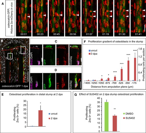
Stump Osteoblasts Proliferate in an FGF-Dependent Manner (A) osteocalcin:GFP+ osteoblasts are capable of dividing after amputation. Frames from a movie obtained from an osteocalcin:GFP; Histone 2a-mCherry transgenic fish fin ray at 1 dpa. Times are indicated (hours:minutes). Scale bar, 20 μm. (B–D) GFP+ and GFP- stump osteoblasts incorporate BrdU (1 hr pulse) in osteocalcin:GFP fish at 1 dpa. (B) Overview. (C) A BrdU+ osteoblast that is Zns 5+ but negative for GFP (arrow) (n = 4 fish, 37/44 sections). Also note the GFP+ cell located distally to the amputation plane (asterisk). (D) A BrdU+ osteoblast that is positive for Zns 5 and GFP (arrowhead) (n = 4 fish, 5/17 sections). Scale bars, 50 μm (B) and 10 μm (C and D). (E) Proliferation of Zns 5+ cells in the distal stump (0–350 μm proximal to the amputation plane at 2 dpa or equivalent area in uncut fins) is highly upregulated at 2 dpa as indicated by BrdU incorporation (5 mM for 9 hr). Error bars represent SEM. Student′s t test, p = 0.021 (nuncut = 3 fish with 31 sections; n2 dpa = 3 fish with 43 sections). (F) Quantification of the proliferative response of stump osteoblasts at 3 dpa. Average number +SEM of BrdU+ Zns 5+ cells (45 min BrdU pulse) per section is shown in bins of 175 μm along the proximal-distal axis (n = 5 fish each, uncut: 214 sections; 3 dpa: 284 sections). Student′s t test, *p < 0.05, ***p < 0.001. (G) Inhibition of FGF signaling by the FGFR1 inhibitor SU5402 significantly downregulates proliferation in distal stump osteoblasts at 2 dpa, as determined by BrdU incorporation (5 mM for 9 hr). Error bars represent SEM. Student′s t test, **p = 0.0013 (nDMSO = 4 fish with 43 sections; nSU5402 = 4 fish with 40 sections). See also Figure S2. |
| Genes: | |
|---|---|
| Fish: | |
| Condition: | |
| Anatomical Term: | |
| Stage: | Adult |
Reprinted from Developmental Cell, 20(5), Knopf, F., Hammond, C., Chekuru, A., Kurth, T., Hans, S., Weber, C.W., Mahatma, G., Fisher, S., Brand, M., Schulte-Merker, S., and Weidinger, G., Bone Regenerates via Dedifferentiation of Osteoblasts in the Zebrafish Fin, 713-724, Copyright (2011) with permission from Elsevier. Full text @ Dev. Cell

