
Fig. 3
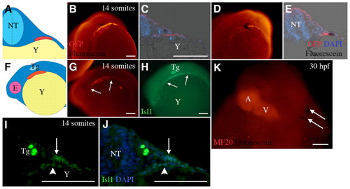
Cells that contribute to the arterial pole remain outside the early heart tube. (A) Diagram of a transverse section of a 14-somite embryo, with dorsal to the top, showing the neural tube (NT, light blue) with differentiating myocardium (red) in the ALPM overlying the yolk (Y, yellow). (B) Whole-mount 14-somite embryo in caudal view, dorsal to top. Labeled cells from grid zone F (black) were observed in the splanchnic mesoderm of Tg(cmlc2::GFP) zebrafish that expresses GFP in differentiated cardiomyocytes (orange). (C) Transverse section oriented as in A showing labeled cells (black) relative to differentiating myocardium (red). (D,E) Cells from grid zone B (black) can be seen in the differentiated myocardium (red) at 14 somites in whole mount (D) and in section (E). (F) Diagram of a 14-somite embryo from the left side, dorsal to top, showing the position of the cardiac progenitors (red), the eye (E, pink), the trigeminal placode (Tg, light blue) and the yolk (yellow). (G,H) A 14-somite embryo oriented as in F, showing Isl1 (H, green) and cardiomyocytes (G, orange). The trigeminal placode and the non-differentiating portion of the heart field are Isl1 positive. Arrows indicate the anterior and posterior extent of cmlc2::GFP or Isl1 signal in G and H, respectively. (I,J) Transverse sections of embryos at the 14-somite stage at the level of the trigeminal placode. Isl1 (green) can be barely detected in the nuclei (blue) of differentiating myocardium (arrowhead) but more brightly in the nuclei of cells in the adjacent splanchnic mesoderm (arrow). (K) Dorsal view of a 30 hpf zebrafish. Cells uncaged from grid zone F at the 7-somite stage (black, arrows) can be seen at some distance from the heart tube (orange). A, atrium; V, ventricle. Scale bars: 10 μm. |