
Fig. S9
- ID
- ZDB-FIG-110429-27
- Publication
- Fujita et al., 2011 - Assembly and patterning of the vascular network of the vertebrate hindbrain
- Other Figures
- All Figure Page
- Back to All Figure Page
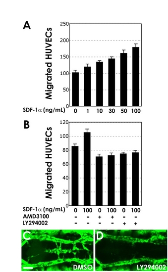
PI3K signaling is required for HUVEC migration in vitro and for PHBC migration in vivo. (A) SDF-1α-induced dose-dependent migration of HUVECs after a 5 hour incubation, measuring the number of transwell-migrated cells. (B) HUVEC transwell migration in response to 100 ng/ml SDF-1α (column 2) and after pretreatment with either 1 mM AMD3100 (column 4) or 10 mM LY294002 (column 6). Shown (A,B) are the mean (with s.e.m.) obtained by measuring eight randomly selected fields of transwell-migrated cells. (C,D) Confocal images of PHBCs and BA in the hindbrain of 32 hpf Tg(fli1a:EGFP)y1 animals treated from 24-32 hpf with either control DMSO carrier (C) or the PI3K inhibitor LY294002 (D). Dorsal view, rostral to the left. Scale bar: 50 μm. |