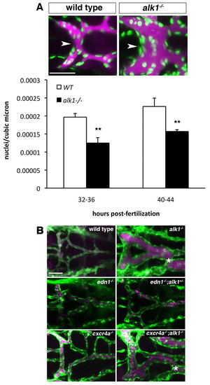
Vasodilation contributes to increased vessel caliber in alk1 mutants. (A) Qtracker 655 non-targeted quantum dots were injected into the common cardinal vein of wild-type and alk1 mutant Tg(fli1a:nEGFP)y7 embryos. Images are two-dimensional confocal projections, dorsal views, anterior leftwards. Embryos aged 32-36 hpf are shown. Endothelial cell nuclei are green; quantum dots are magenta. Scale bar: 50 μm. Endothelial cell density in the BCA (white arrowhead) was calculated from these micrographs by dividing the number of nuclei by the vessel volume. Data represent mean±s.e.m. (n=5-7 independent samples) and were analyzed using Student′s t-test, **P<0.001. (B) Analysis of the contribution of edn1 loss and cxcr4a upregulation to AVM development. AVM development in alk1-/- embryos (asterisk) is not phenocopied by edn1-/- embryos even in the presence of heterozygous levels of alk1 (edn1-/-;alk+/-). cxcr4a-/- embryos do not exhibit AVMs, whereas cxcr4a-/-;alk1+/- embryos invariably develop AVMs, demonstrating that cxcr4a upregulation is not necessary for AVM development in alk1 mutants. Two-dimensional confocal projections of Tg(fli1a:EGFP)y1;Tg(gata1:dsRed)sd2 or Tg(kdrl:GFP)la116;Tg(gata1:dsRed)sd2 embryos. Endothelial cells are green; erythrocytes are magenta. Dorsal views, anterior leftwards. Scale bar: 50 μm.
|
