
Fig. 5
- ID
- ZDB-FIG-110405-15
- Publication
- Choi et al., 2011 - Aplexone targets the HMG-CoA reductase pathway and differentially regulates arteriovenous angiogenesis
- Other Figures
- All Figure Page
- Back to All Figure Page
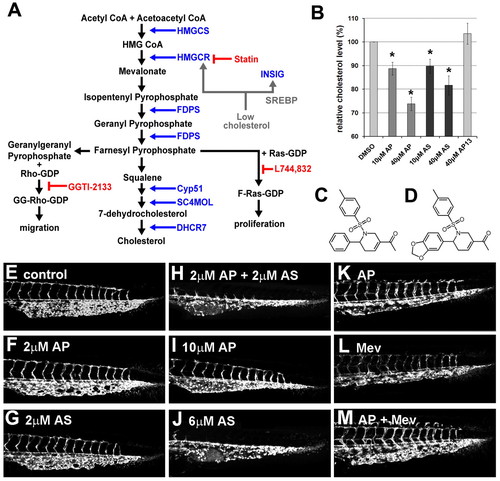
Aplexone affects the HMGCR pathway. (A) Simplified schematic diagram of the HMGCR pathway. Enzymes involved in the pathway are shown in blue. Feedback loop is indicated in gray. Chemical inhibitors of the pathway are indicated in red. HMGCS, 3-hydroxy-3-methylglutaryl-CoA synthase; HMGCR, HMG CoA reductase; FDPS, farnesyl diphosphate synthetase; Cyp51, lanosterol 14 α-demethylase; SC4MOL, sterol-C4-methyl oxidase-like; DHCR7, 7-dehydrocholesterol reductase; INSIG, insulin induced gene 1; SREBP, sterol regulatory element binding proteins. (B) Relative cholesterol levels in embryos treated with 0.4% DMSO as a control (DMSO), aplexone (AP, structure shown in C), atorvastatin (AS) or a non-functional aplexone analog (AP13, structure shown in D). Both aplexone and atorvastatin reduce cholesterol levels significantly (*P<0.05). Data are mean±s.e.m. (E-J) Aplexone and atorvastatin have a synergistic effect on caudal vein angiogenesis. Embryos were treated with the chemicals indicated beginning at 10 hpf and caudal vein images were taken at 48 hpf. (K-M) Mevalonate reverses the effect of aplexone on caudal vein plexus formation. Caudal vein plexus of embryos treated with 5 μM aplexone beginning at 2 hpf (K), injected with 1 nl of 2 M mevalonate (L) or injected with mevalonate followed by aplexone treatment (M). |