
Fig. 2
- ID
- ZDB-FIG-110322-12
- Publication
- Mickoleit et al., 2011 - Regulation of hub mRNA stability and translation by miR430 and the dead end protein promotes preferential expression in zebrafish primordial germ cells
- Other Figures
- All Figure Page
- Back to All Figure Page
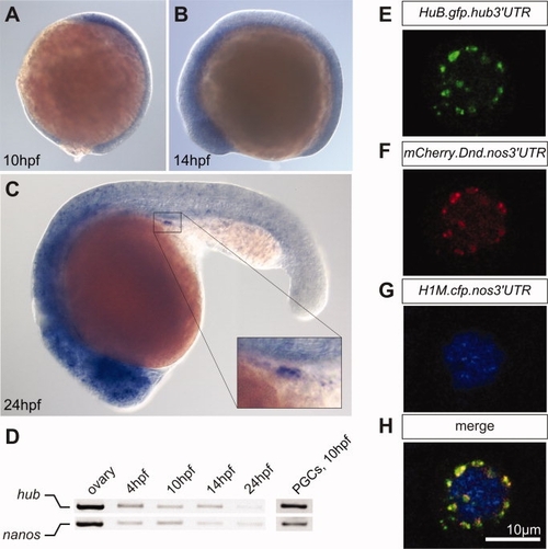
Figure 2. hub mRNA is expressed in the head and in PGCs of zebrafish embryos and HuB protein is localized to perinuclear granules in the germ cells. A–C: Whole-mount in situ hybridization of wild type zebrafish embryos using an antisense probe directed against zebrafish hub RNA at the indicated developmental stages. hub mRNA is ubiquitously expressed in 10-hpf (A) and in 14-hpf embryos (B). In 24-hpf embryos, hub RNA is expressed in the head and in PGCs (inset) (C). D: The expression of nanos and hub mRNA determined by RT-PCR. Ovary cDNA, cDNA of embryos of the indicated developmental stages, and cDNA of germ cells isolated from 10hpf embryos were used as templates. E–H: Confocal microscopy images of a PGC in a 6hpf embryo showing the sub-cellular localization of the HuB-GFP fusion protein. The HuB fusion protein is localized to granular structures around the nucleus (E), which are co-labeled with the mCherry-Dnd fusion protein (F). The nucleus is marked by the CFP-histone protein (G) and the merged image is shown in H. |
| Genes: | |
|---|---|
| Fish: | |
| Anatomical Terms: | |
| Stage Range: | Sphere to Adult |