
Fig. 3
- ID
- ZDB-FIG-110321-5
- Publication
- Wibowo et al., 2011 - Compartmentalized Notch signaling sustains epithelial mirror symmetry
- Other Figures
- All Figure Page
- Back to All Figure Page
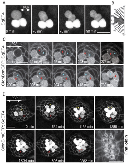
Orientation of UHCP divisions and planar cell inversion. (A) A 90-minute series of confocal images of a regenerating SqET4 neuromast revealing two GFP-positive hair cells aligned along the axis of planar polarity (double-headed arrow). An UHCP develops at the upper edge of the neuromast. Over the next hour, this UHCP commences mitosis and divides obliquely into a pair of hair cells. (B) Quantification of the angle of division of UHCPs in 23 neuromasts, which indicates that the majority of UHCP divide obliquely respect to the axis of planar polarity of the epithelium. (C) A 12-hour series of confocal images of a regenerating neuromast of a double-transgenic Tg[Cldnb:mGFP;SqET4] larva revealing two GFP-positive hair cells at the lower aspect of the image, and a UHCP at the top. At 14 minutes into the time-series, this UHCP divides into two hair cells, each identified with a dot (red and blue). At 264 minutes after cytokinesis (2762), the sibling hair cells begin to rotate around their contact point within the plane of the epithelium, which locally breaks the line of mirror symmetry (this is most evident at 306 minutes into the time series). A complete inversion of the hair-cell pair eventually reforms the line of mirror symmetry and precise realigns the cells along the axis of planar polarity of the neuromast (double-headed arrow in the first panel). (D) A 44-hour series of confocal images of a regenerating neuromast of a double-transgenic Tg[Cldnb:mGFP;SqET4] larva reveals plasma-membrane GFP-positive supporting cells and cytoplasmatic GFP-positive hair cells and UHCPs. Two prospective UHCPs were identified retrospectively by playing the time series backwards, and each labeled with either a red or yellow dot. Their position was followed over time, showing that prospective UHCPs moved into the dorsal (yellow) and ventral (red) polar compartments, where they became UHCPs (weak cytoplasmatic GPF). Each UHCP eventually divided into a pair of hair cells. The last panel of the image is an actin staining, showing the resulting planar polarization of the epithelium. The axis of planar polarity of the neuromast is indicated by a double-headed arrow in the first panel. Scale bars: 10 μm. |