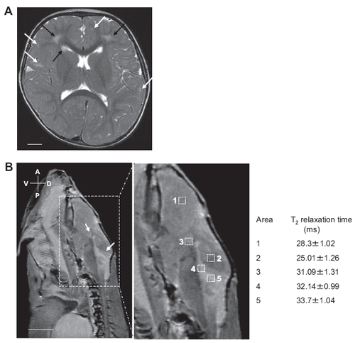
Comparable MR presentations of RNase T2-deficient human and zebrafish. (A) Axial T2-weighted cerebral MR image of a patient with RNASET2-deficient cystic leukoencephalopathy. The image indicates multifocal white matter abnormalities adjacent to the ventricles (black arrows) and in deep white matter (white arrows). (Scale bar: 1 cm). (B) T2 relaxation time measurement of the brain of a mutant zebrafish containing nullizygous mutation in rnaset2 gene. Sagittal MR image of the brain of an adult AO127 mutant zebrafish indicate the presence of lesions adjacent to the ventricles (white arrows) and were of similar intensity as that of CSF present in ventricles. (Boxed area, enlarged at right.) Areas were selected for T2 relaxation time measurements within healthy brain region (1, 2), in ventricles (4), and in lesion outside (3) or adjacent to ventricle (5). The lesions adjacent to ventricles have similar T2 relaxation time as that of ventricles, suggesting that these lesions may be filled with solution similar to CSF.
|
