
Fig. 4
- ID
- ZDB-FIG-110121-42
- Publication
- Bernardi et al., 2010 - Characterization of the Regulatory Region of the Zebrafish Prep1.1 Gene: Analogies to the Promoter of the Human PREP1
- Other Figures
- All Figure Page
- Back to All Figure Page
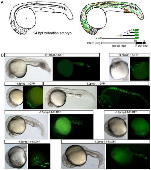
Transient expression of zebrafish prep1.1:GFP promoter constructs in 24 hpf zebrafish embryos. A) Schematic drawing of a 24 hpf zebrafish embryo, in which the main anatomical features are shown. On the same drawing, the transient expression patterns of all the zebrafish promoter contructs used for the analysis are reported: each dot corresponds to a group of GFP-positive cells, whereas each color is related to different promoter region (see map below). This pattern is the result of dozens of observations (see Table 3) manually annotated after each microinjection experiment and stereomicroscope observations. B) For each construct (indicated in the stripe at the top of each figure), a representative image of its transient expression in vivo is shown. Sometimes the entire embryo is shown (in the lateral view), sometimes only the anterior region (in frontal or lateral view). e, eye; Et, intronic enhancer; h, hindbrain; m, midbrain; n, notochord; sc, spinal chord; t, telencephalon; TSS, Transcription Start Site; y, yolk; ye, yolk extension. |