
Fig. 3
- ID
- ZDB-FIG-110112-56
- Publication
- Dolez et al., 2011 - Laminins, via heparan sulfate proteoglycans, participate in zebrafish myotome morphogenesis by modulating the pattern of Bmp responsiveness
- Other Figures
- All Figure Page
- Back to All Figure Page
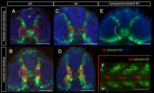
pSmad expression is precisely regulated in pioneers and their precursors in wild-type, sly and cyclopamine-treated embryos. (A-E) Transverse sections of 18-20 s stage wild-type (A,B), sly (C,D) and cyclopamine-treated embryos (E) at the level of adaxial cell pre-stacking (A,C,E) and stacking (B,D). Dorsal is upwards. F59 (in red) labels adaxial cells and nuclear pSmad labelling is in green; nuclei are counterstained with DAPI (in blue). (F) Lateral view (projection of 11 confocal sections, 0,6 μm z-step) of a 18-20 s stage wild-type embryo, at the trunk level. Anterior is on the left, dorsal is upwards. 4D9 (in red) labels pioneers and nuclear pSmad labelling is in green. n, notochord; nt, neural tube. Scale bar: 50 μm. |