|
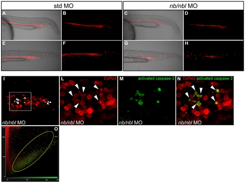
dsRed+ erythroid cells are dramatically reduced at 28–30 hpf. A–H. Tg(gata1:dsRed) embryos injected with std MO (A, B, E, F) and nb/nbl MO (C, D, G, H) were examined by confocal microscopy between 24–30 hpf. Fluorescent images (B, D, F, H) were merged with bright field images (A, C, E, G). In nb/nbl morphants at 24–26 hpf red fluorescent erythroid cells are present within the ICM (C, D) but at 28–30 hpf the overall fluorescence of nb/nbl morphants appears strongly reduced (G, H). I–N. Whole-mount double immunofluorescence on Tg(gata1:dsRed) nb/nbl morphants at 28–30 hpf to detect caspase-3 activation (green) and DsRed (Red). Single optical section of nb/nbl morphants obtained by confocal microscopy (20x magnification, I). I. White spots, indicating double positive cells, have been pseudocoloured according to the region of interest (ROI1 in O). The sub-image area, shown in detail in panels L–N, is highlighted by the white box. L–N. Insets of single channel fluorescent images (L, M) and merge (N) are shown. Activated caspase-3 overlaps with some dsRed+ cells (white arrowheads, L). O. Fluorogram shows the degree of colocalization between red signals (dsRed) and green signals (activated caspase-3); colocalization is indicated by the region of interest (ROI1).
|