
Fig. 2
- ID
- ZDB-FIG-101223-26
- Publication
- Lee et al., 2010 - The habenula prevents helpless behavior in larval zebrafish
- Other Figures
- All Figure Page
- Back to All Figure Page
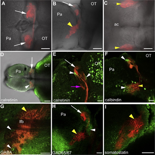
Characterization of KillerRed-Expressing Habenula Afferents (A–C) Forebrain of a KR11 zebrafish in dorsal (A), lateral (B), and ventral (C) views. KillerRed is expressed in axons that innervate the habenula (arrows). Cell bodies of KillerRed-expressing neurons (arrowheads) are in the ventral forebrain (B), in a lateral position (C). (D) Dorsal view of a 2-week-old fish showing calretinin label in restricted habenula neuropils (arrowhead). (E) Sagittal section showing calretinin label in two dorsal habenula neuropils (arrowheads). (F) Lateral view (projection) showing calbindin label in KillerRed-positive neurons (white arrowhead). (G) Lateral view showing rare GABA-positive neurons (arrowheads) in the KillerRed-expressing population. The lateral forebrain bundle is visible in this optical section. (H) Projection of the left side, showing GAD65/67 label in neurons (white arrowhead) dorsal to the KillerRed cluster. (I) Optical section, lateral view, showing lack of somatostatin label in neurons expressing KillerRed. The following abbreviations are used: Pa, pallium; OT, optic tectum; ac, anterior commissure; lfb, lateral forebrain bundle. Scale bars represent 50 μm in (A)–(D) and 20 μm in (E)–(I). Yellow arrowheads indicate KillerRed-expressing cells; white arrows indicate the habenula; pink arrow indicates ventral habenula. Anterior is to the left in all cases. Fish in (F)–(H) are 3 weeks old; fish in (A)–(C), (E), and (I) are 4 weeks old. |