
Fig. 5
- ID
- ZDB-FIG-101208-26
- Publication
- Tu et al., 2010 - Clonal analyses reveal roles of organ founding stem cells, melanocyte stem cells and melanoblasts in establishment, growth and regeneration of the adult zebrafish fin
- Other Figures
- All Figure Page
- Back to All Figure Page
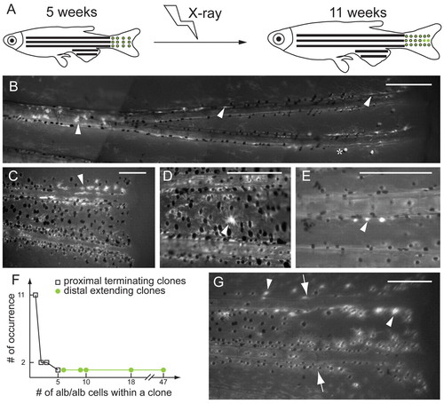
X-ray induction of albino clones in mature stage of albino/+; Tg(fTyrp>GFP)j900 fish reveals growth potential of MSCs and melanoblasts. (A) At 5 weeks, fish are exposed to 920 Rads of X-rays to induce loss-of-heterozygosity at the albino locus. albino melanocytes are revealed by expression from the fTyrp>GFP transgene upon examination 6 weeks later. (B,C) Fluorescence images of two examples of clone class that extends distally. Clones of albino cells begin in the interior of the fin and extend distally to the edge of the fin. Asterisk denotes the white cells, which reflect incident light, rather than fluorescence. (D,E) Fluorescence images of two examples of clone class located in proximal position without extending to distal edge of fin. (F) Graph shows occurrence number of clones versus the number of albino melanocytes for each clone class. (G) Regeneration of fin carrying a distal-extending clone. (B-E,G) Arrowheads indicate albino melanocytes, and (G) arrows indicate the amputation plane. Scale bars: 0.2 mm. |