
Fig. S7
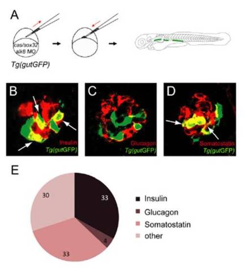
Mosaic knockdown of alk8 preferentially generates additional Insulin and Somatostatin-expressing cells. (A) Schematic diagram of the cell transplantation protocol. Tg(gutGFP) donor embryos were injected with cas/sox32 mRNA and alk8 splice-blocking MO before transplantation into wild-type hosts. (B–D) Confocal images of hosts containing Tg(gutGFP) donor cells (green) and stained for Insulin (B), Glucagon (C), or Somatostatin (D) (red) at 72 hpf. (E) Proportional distribution of hormone content of alk8 morphant cells located in the islet. The percentages were calculated by counting the number of donorderived hormone-expressing cells and dividing it by the total number of the donor cells in the islet (total number of cells counted = 306). Host embryos were stained for Insulin or Glucagon/Somatostatin; because the sum of the percentages of Insulin, Glucagon, and Somatostatin-expressing cells did not reach 100%, the data indicate that some donor cells differentiated into other endocrine cell types (e.g., ε- or γ-cells), or nonendocrine cells. |