
Fig. 2
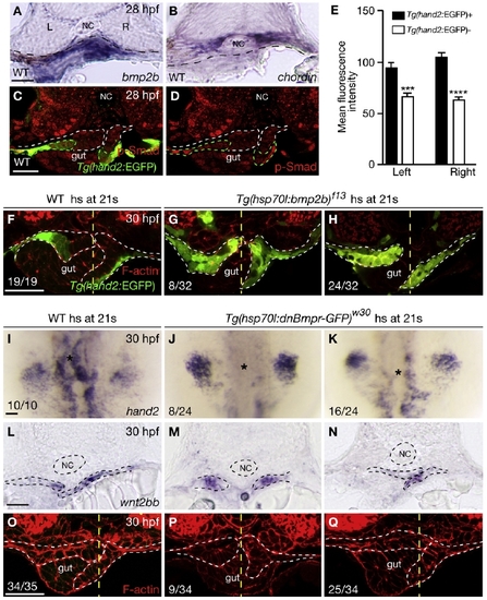
Bmp Signaling Regulates hand2 Expression in the LPM (A and B) Within the gut-looping region of wild-type embryos, bmp2b (A) is expressed in the LPM, whereas chordin (B) is expressed in the ventral portion of the somites. Dashed lines mark the somite/LPM boundary. (C) Wild-type embryo stained for GFP (green) and phosphorylated Smad1/5/8 (red). (D) The same embryo as shown in (C) but with red channel only. Dashed lines outline the LPM. (E) Fluorescence intensity of the phosphorylated Smad staining in the LPM cells (mean ± SEM). Forty Tg(hand2:EGFP)-expressing cells and 40 Tg(hand2:EGFP)-nonexpressing cells from seven wild-type embryos were analyzed. Asterisks indicate statistical significance: ***p < 0.001; ****p < 0.0001. (F–H) Wild-type and Tg(hsp70l:bmp2b)f13 embryos were heat shocked at the 21-somite stage, and stained for GFP (green) and phalloidin (red) at 30 hpf. White dashed lines outline the LPM. Yellow dashed lines mark the midline. Tg(hand2:EGFP) was expressed mostly in the ventral half of the LPM in wild-types (F), but was expressed throughout the LPM in the gut-looping region in Tg(hsp70l:bmp2b)f13 embryos (G and H). Eight of 32 Tg(hsp70l:bmp2b)f13 embryos examined exhibited no gut looping (G); the remaining 24 exhibited leftward gut looping (H). (I–K) Compared to wild-type control (I), hand2 expression in the LPM (asterisks) was decreased (K) or absent (J) in Tg(hsp70l:dnBmpr-GFP)w30 embryos after heat shock. (L–N) Expression of wnt2bb in the LPM was present in wild-type and Tg(hsp70l:dnBmpr-GFP)w30 embryos after heat shock. Dashed lines outline the notochord and LPM. (O–Q) Phalloidin staining of wild-type control (O) and Tg(hsp70l:dnBmpr-GFP)w30 (P and Q) embryos. When heat shock was applied at the 21-somite stage, 9 of 34 embryos exhibited no gut looping (P); the remaining 25 exhibited leftward gut looping (Q). White dashed lines outline the LPM. Yellow dashed lines mark the midline. All images, except (I)–(K), are transverse sections, dorsal to the top. (I)–(K) are dorsal views, anterior to the top. L, left; NC, notochord; R, right. The scale bars represent 40 μm. See also Figure S1. |
| Genes: | |
|---|---|
| Antibody: | |
| Fish: | |
| Condition: | |
| Anatomical Terms: | |
| Stage Range: | Prim-5 to Prim-15 |
| Fish: | |
|---|---|
| Condition: | |
| Observed In: | |
| Stage: | Prim-15 |
Reprinted from Developmental Cell, 18(6), Yin, C., Kikuchi, K., Hochgreb, T., Poss, K.D., and Stainier, D.Y., Hand2 Regulates Extracellular Matrix Remodeling Essential for Gut-Looping Morphogenesis in Zebrafish, 973-984, Copyright (2010) with permission from Elsevier. Full text @ Dev. Cell