
FIGURE
Fig. 2
- ID
- ZDB-FIG-101124-18
- Publication
- Tschopp et al., 2010 - Funduscopy in adult zebrafish and its application to isolate mutant strains with ocular defects
- Other Figures
- All Figure Page
- Back to All Figure Page
Fig. 2
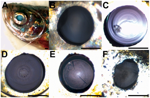
Images of zebrafish eyes. (A) Overview of anaesthetized fish with cover glass, (B) shows a normal, transparent lens, (C–F) examples of opaque lenses (cataracts): (C) small inclusion are present in an otherwise normal lens, (D) massive cataract, (E) including cracks in the lens, and (F) membranous cataract. Scale bar: 500 μm. |
Expression Data
Expression Detail
Antibody Labeling
Phenotype Data
| Fish: | |
|---|---|
| Observed In: | |
| Stage: | Adult |
Phenotype Detail
Acknowledgments
This image is the copyrighted work of the attributed author or publisher, and
ZFIN has permission only to display this image to its users.
Additional permissions should be obtained from the applicable author or publisher of the image.
Full text @ PLoS One