
Fig. 5
- ID
- ZDB-FIG-101117-27
- Publication
- Lee et al., 2009 - Hypoxia-induced pathological angiogenesis mediates tumor cell dissemination, invasion, and metastasis in a zebrafish tumor model
- Other Figures
- All Figure Page
- Back to All Figure Page
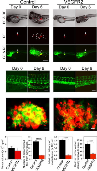
Inhibition of tumor cell invasion, dissemination and metastasis by VEGFR2 morpholinos. (A and D) VEGFR2 specific morpholinos and control morpholinos were injected into the blastoma of 1 h post-fertilization at 1–4-cell stages. DiI-labeled T241-VEGF tumor cells were implanted in the perivitelline space of 48 h post-fertilization embryos and tumor cell invasion, dissemination and metastasis were detected at days 0 and 6 post-injection. White arrowheads indicate disseminated tumor foci. (Scale bar, 500 μm.) (B and E) High-resolution micrographs of A and D, respectively to visualize single metastatic tumor cells in the trunk regions. (Scale bar, 100 μm.) (C and F) Representative 3-D micrographs of confocal images of tumors (red) and tumor vasculatures (green). (Scale bar, 10 μm.) (G) Quantification of tumor volume (n = 12/group). (H) Quantification of numbers of disseminated tumor foci (n = 12/group). (I) Averages of maximal distances of metastatic foci (n = 12/group). (J) Quantification of tumor vessel density relative to tumor size (n = 7/group). Data are represented as mean ± SEM. |