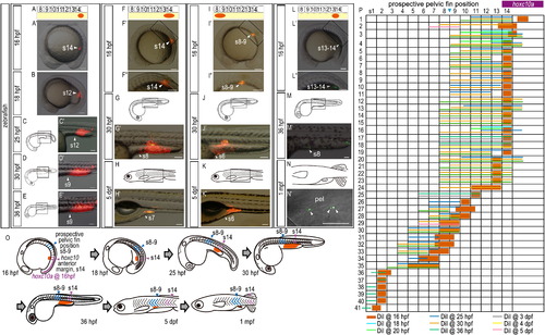
FIGURE

Fig. 1

ID
ZDB-FIG-101108-11
Publication
Murata et al., 2010 - Allometric growth of the trunk leads to the rostral shift of the pelvic fin in teleost fishes
Other Figures
All Figure Page
Back to All Figure Page
Fig. 1

 Positional relationships between the body trunk and the lateral plate mesoderm on the yolk change when the trunk-tail protrudes from the yolk during zebrafish development. (A-N) Cell lineage analysis in zebrafish embryos. Locations of DiI-labeled cells (A-K) and quantum dot-labeled cells (L-N) are indicated by arrowheads. (A, F, I, L) Schematic diagrams showing the locations of DiI or quantum dot (red) application. (A′, F′, I′, L′) DiI or quantum dot was applied to the lateral plate mesoderm cells opposite somites 14 (A′, F′, L′) and 8-9 (I′) in 16 hpf zebrafish embryos. (F3, I3, L3) Higher magnification of the labeled regions shown in (F′), (I′) and (L′), respectively. (C, D, E, G, H, J, K, M, N) Schematic diagrams of zebrafish embryos at 25 hpf (C), 30 hpf (D, G, J), 36 hpf (E, M), 5 dpf (H, K) and 1 mpf (N). (O) Schematic diagram summarizing the lineage of labeled cells. Lateral plate mesodermal cells opposite somite 14 at 16 hpf (red circles), next to the anterior margin of hoxc10a expression in the spinal cord (purple arrowheads), were found at the level of somites 8-9 by 36 hpf and persisted in the same region at 5 dpf, where pelvic fin buds appear (blue arrowheads). (P) Cell lineage analysis in zebrafish embryos. Numbers above each column indicate the opposite somite levels. Expression of hoxc10a in the spinal cord at 16 hpf is indicated by the purple bar. Sample numbers are shown at left. Bold red bars indicate the width of the area labeled with DiI at 16 hpf in each experiment. Light blue, light green, blue, orange, green, grey, yellow and pink bars indicate the width of the area labeled with DiI at 18, 20, 25, 30, 36 hpf, 3 dpf, 4 dpf and 5 dpf, respectively. Note that the DiI-labeled cells around the anterior border of hoxc10a expression in the spinal cord of embryos at 16 hpf were identified in the prospective pelvic fin position by 30 hpf, and continued to be in the same position at 36 hpf, 3 dpf and 5 dpf. Small numbers of lateral plate cells close to the body trunk shift caudally, as they extend together with the body trunk. Scale bars: 100 μm.

Expression Data

Expression Detail
Antibody Labeling
Phenotype Data

Phenotype Detail
Acknowledgments
This image is the copyrighted work of the attributed author or publisher, and ZFIN has permission only to display this image to its users. Additional permissions should be obtained from the applicable author or publisher of the image.

Reprinted from Developmental Biology, 347(1), Murata, Y., Tamura, M., Aita, Y., Fujimura, K., Murakami, Y., Okabe, M., Okada, N., and Tanaka, M., Allometric growth of the trunk leads to the rostral shift of the pelvic fin in teleost fishes, 236-245, Copyright (2010) with permission from Elsevier. Full text @ Dev. Biol.