
Fig. 1
- ID
- ZDB-FIG-101102-3
- Publication
- Bagnat et al., 2010 - Cse1l Is a Negative Regulator of CFTR-Dependent Fluid Secretion
- Other Figures
- All Figure Page
- Back to All Figure Page
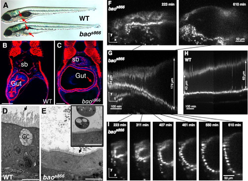
baos866 Mutants Undergo a Dramatic and Rapid Expansion of the Gut Lumen between 96 and 120 Hours Postfertilization (A) Bright-field image of 120 hr postfertilization (hpf) wild-type (WT) and baos866 mutant larvae. Arrows point to the edges of the gut lumen. (B and C) Confocal images of cross-sections of 120 hpf WT (B) and baos866 mutant (C) larvae. The arrow in (C) points to a delaminating cell. Red is F-actin and blue is DAPI; sb indicates swim bladder. Scale bars represent 50 μm. (D and E) Dramatic shortening of microvilli (arrows) in baos866 mutant enterocytes observed by transmission electron microscopy at 120 hpf. Inset in (E) shows remnants of apoptotic cells found in the lumen. GC indicates goblet cell. Scale bars represent 10 μm. (F–I) Rapid expansion of the gut lumen in baos866 mutants expressing histone 2A:GFP. (F) Still images (lateral views) from a baos866 mutant selective plane illumination microscopy (SPIM) recording between 96 and 120 hpf showing first and last frames. (G and H) Kymographs from baos866 mutant (G) and WT (H) gut. Lumen expansion in the mutant occurred in ~200 min; no cell division was observed. (I) Still images (lateral views) from the baos866 mutant SPIM recording corresponding to the kymograph shown in (G). |
| Gene: | |
|---|---|
| Fish: | |
| Anatomical Term: | |
| Stage: | Day 4 |
| Fish: | |
|---|---|
| Observed In: | |
| Stage: | Day 5 |