FIGURE

Fig. 6

ID
ZDB-FIG-101025-3
Publication
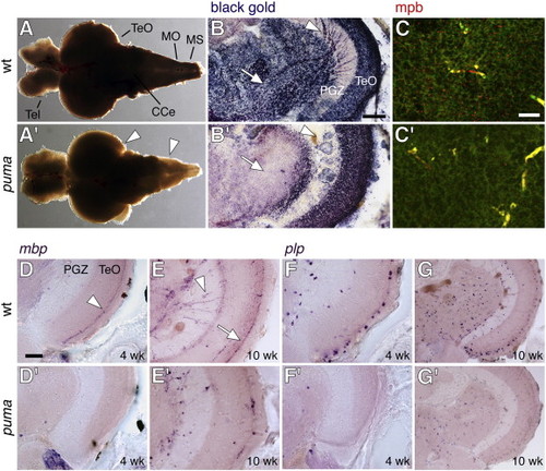
Larson et al., 2010 - Defective adult oligodendrocyte and Schwann cell development, pigment pattern, and craniofacial morphology in puma mutant zebrafish having an alpha tubulin mutation
Other Figures
All Figure Page
Back to All Figure Page
Fig. 6

Deficient central nervous system myelination in puma mutant adults and juveniles. (A, A′) Decreased opacity of adult brain from puma as compared to wild-type; this difference is especially notable at the lateral edges and posteriorly (arrowheads). Anterior to the left. Tel, telencephalon; TeO, tegmentum opticum; CCe, corpus cerebelli; MO, medulla oblongata; MS, medulla spinalis. (B, B′) Black Gold II staining for myelin in transverse sections of wild-type and puma mutant adults. Myelin is stained blue-black whereas other tissue is purple-red. Reduced staining in puma is evident to some degree within the optic tectum (TeO) but is more pronounced in ventral–medial regions (arrow), internal to the periventricular grey zone (PGZ). Radially oriented black gold staining (arrowhead) also is largely absent from the PGZ itself of the puma mutant. (C, C′) Antiserum against zebrafish mbp (red) stains numerous punctate foci in the ventral–medial brain of wild-type but far fewer in the corresponding region of puma mutants. Green, background tissue fluorescence. Yellow, fluorescence of blood vessels in green and red channels. (D, D′–G′, G) In situ hybridization for mbp and plp in juveniles and adults (13 SSL, 24 SSL). (D, D′) In juveniles, staining for mbp is pronounced in a band (arrowhead) near the outer surface of the optic tectum (TeO) in wild-type (D) but is not apparent in puma (D′). (E, E′) In adults, abundant mbp transcripts are found radially in the PGZ (arrowhead) and at the outer surface of the optic tectum (arrow) in wild-type but are found at lower levels in puma. (F, F′) In juveniles, plp staining is detected in individual cell bodies both interiorly (arrowhead) and near the tectum surface (arrow) of wild-type but there are far fewer of these cells in puma mutants. (G, G′) In adults, numerous plp-expressing cell bodies are observed in wild-type, whereas in puma mutants there are fewer cells and those present are less densely stained. Scale bars: in B, 100 μm; in C, 20 μm; in D, 100 μm for D–G.

Expression Data
Genes:
Fish:
Anatomical Terms:
Stage Range: Days 30-44 to Adult

Expression Detail
Antibody Labeling
Phenotype Data
Fish:
Observed In:
Stage Range: Days 30-44 to Adult

Phenotype Detail
Acknowledgments
This image is the copyrighted work of the attributed author or publisher, and ZFIN has permission only to display this image to its users. Additional permissions should be obtained from the applicable author or publisher of the image.

Reprinted from Developmental Biology, 346(2), Larson, T.A., Gordon, T.N., Lau, H.E., and Parichy, D.M., Defective adult oligodendrocyte and Schwann cell development, pigment pattern, and craniofacial morphology in puma mutant zebrafish having an alpha tubulin mutation, 296-309, Copyright (2010) with permission from Elsevier. Full text @ Dev. Biol.