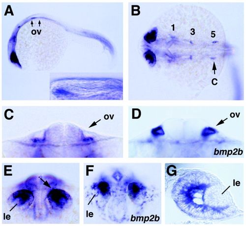
smad1 and bmp2b expression in eyes and ears. All embryos are shown at 36 hr after fertilization. A:smad1, lateral view; the anterior and posterior border of the otic vesicles (ov) is indicated by arrows. The inset shows a magnification of the indicated region with the otic vesicle. smad1 is expressed in mesenchyme ventral of the anterior region of the otic vesicle. B:smad1, dorsal view on head. Numbers 1, 3, and 5 mark the three bilateral expression domains associated with rhombomeres 1, 3, and 5. The position of the optical cross section shown in (C) is indicated. C, D:smad1(C) and bmp2b (D), optical cross section at level of otic vesicles (ov). smad1 is expressed in mesenchyme ventral of the vesicle, bmp2b in the vesicle epithelium. E, F:smad1 (E) and bmp2b (F), anterior view on head; smad1 and bmp2b are expressed in a subset of retinal cells close to the developing lens (le). In (E), the position of the section shown in (G) is indicated by an arrow. G:smad1, section through eye vesicle of embryo shown in (E); smad1 is strongly expressed in presumptive ganglion cells close to the lens, whereas expression in outer regions of the retina is rather weak. Abbreviations: le, lens; ov, otic vesicle.
|
