
Fig. 7
- ID
- ZDB-FIG-100804-11
- Publication
- Roman et al., 2002 - Disruption of acvrl1 increases endothelial cell number in zebrafish cranial vessels
- Other Figures
- All Figure Page
- Back to All Figure Page
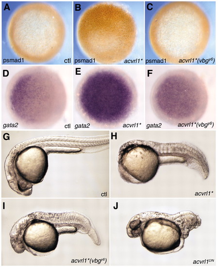
Zebrafish Acvrl1 acts through the Smad1/5/8 pathway in vivo. Shield stage embryos injected at the 1- to 4-cell stage with 5 pg mRNA encoding an activated form of Acvrl1 (acvrl1*) exhibit ectopic Smad1 phosphorylation (B) compared to uninjected controls (A). Expression of gata2 is also upregulated in acvrl1*-injected embryos (E) compared to uninjected controls (D). In contrast, 100 pg acvrl1*(vbgy6) mRNA was ineffective in inducing Smad1 phosphorylation (C), and only minimally effective in inducing gata2 expression (F). When assayed at 24-30 hpf, embryos injected with 1 pg acvrl1* mRNA (H) exhibited strong ventralization compared to uninjected controls (G), whereas those injected with 100 pg acvrl1*(vbgy6) mRNA were less severely ventralized (I). Embryos injected with 100 pg mRNA of a kinase-dead, dominant negative form of acvrl1 (acvrl1DN) exhibit strong dorsalization (J), whereas those injected with 600 pg acvrl1DN(vbgy6) were indistinguishable from wild type (data not shown). (A-F) Animal view; (G-J) lateral view, anterior to the left. |