
Fig. S1
- ID
- ZDB-FIG-100707-40
- Publication
- Rojas-Muñoz et al., 2009 - ErbB2 and ErbB3 regulate amputation-induced proliferation and migration during vertebrate regeneration
- Other Figures
- All Figure Page
- Back to All Figure Page
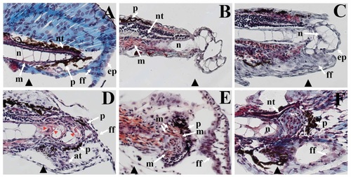
Regenerated structures in zebrafish larvae. (A) Masson’s trichrome staining of the paraffin embedded sections of caudal fins show the cells of the neural tube (nt), notochord (n), muscle fibers (m), as well as pigment cells (p) and epithelium (ep) of the finfold (ff) of an unamputated larval fin. The site of the prospective amputation plane is indicated with a black triangle. (B) 6 hours post injury (hpi), a protrusion from the amputation plane contains vacuolated cells containing very little cytoplasm, a cellular morphology like that of the cells of the notochord. (C) Between 12 and 24 hpi, an epidermal layer (ep), approximately two cells thick, covers the notochord protrusion. Also, finfold tissue (ff) has started to grow distally from the amputation stump. (D) By 3 day post injury (dpi), cells of the finfold now surround the protrusion, and actinotrichia (at, small skeletal filaments of the finfold) are present. Furthermore, cells within the protrusion no longer have the same morphology as cells from the notochord in that they contain more cytoplasm (red staining indicated by red arrowheads). Pigment cells (p, black melanophores) are now present dorsally and ventrally around the protrusion. (E) By 4 dpi, striated muscle fibers surround the protrusion of the fin regenerate. Melanophores (p) now encircle the protrusion, and the architecture of the finfold is very similar to that of the uninjured larval finfold. (F) At 7 dpi, the neural epithelium of the neural tube (nt) is now present in the fin regenerate. Although the basic architecture of the caudal fin is complete, tissue remodeling still appears to be ongoing: there still is a cluster of cells at the distal end of the notochord that have not integrated as a differentiated caudal fin tissue. |
Reprinted from Developmental Biology, 327(1), Rojas-Muñoz, A., Rajadhyksha, S., Gilmour, D., van Bebber, F., Antos, C., Rodríguez Esteban, C., Nüsslein-Volhard, C., and Izpisúa Belmonte, J.C., ErbB2 and ErbB3 regulate amputation-induced proliferation and migration during vertebrate regeneration, 177-190, Copyright (2009) with permission from Elsevier. Full text @ Dev. Biol.