
Fig. S1
- ID
- ZDB-FIG-100628-52
- Publication
- Cerveny et al., 2010 - The zebrafish flotte lotte mutant reveals that the local retinal environment promotes the differentiation of proliferating precursors emerging from their stem cell niche
- Other Figures
- All Figure Page
- Back to All Figure Page
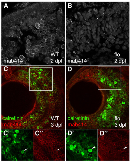
Retinal cells in flo embryos contain disrupted nuclear pores, but still express a marker of neuronal differentiation and function. (A-D″) Nuclear pores and calretinin-positive cells were visualised in wild-type (A,C) and flo (B,D) retinae from 3 dpf by staining with mab414 (grey in A,B, red in C,D) and calretinin (green) sera. C′-D″ are enlargements of the single channels from the boxed regions in C and D, respectively. Calretinin-positive cells (green) from approximately the same retinal position have nuclei ringed uniformly by red mab414 staining (WT, C′,C″, arrow) or have aberrant nuclear pores (flo, D′,D″, arrowhead). |