
Fig. 3
- ID
- ZDB-FIG-100616-108
- Publication
- Kabli et al., 2010 - In vivo magnetic resonance imaging to detect malignant melanoma in adult zebrafish
- Other Figures
- All Figure Page
- Back to All Figure Page
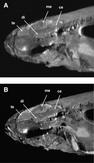
High-resolution images of adult zebrafish at a magnetic field strength of 9.4 T (A) and 17.6 T (B). Slices in sagittal plane were obtained using the rapid acquisition with relaxation enhancement pulse sequence (echo time [TE], 15 ms with effective TE, 33.6 ms; repetition time, 2000 ms; number of scan, 4; total scan time, 8 min). The image resolution is 78 (m and slice thickness is 0.2 mm. Signal-to-noise ratio at 9.4 and 17.6 T was calculated to be 18 and 32, respectively. Image quality improvement is clearly visible at 17.6 T, as many substructures in the brain (e.g., te, telencephalon; di, diencephalon; me, mesencephalon; ce, cerebellum) that were not visible at 9.4 T can be clearly seen at 17.6 T. |