Fig. 3
- ID
- ZDB-FIG-100429-119
- Publication
- Haug et al., 2010 - Visual acuity in larval zebrafish: behavior and histology
- Other Figures
- All Figure Page
- Back to All Figure Page
|
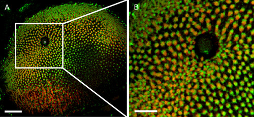
Confocal image of an immunohistochemically labeled 6 dpf zebrafish retina. Red-green double cones and blue cones were each labeled with a specific primary antibody and marked with a secondary antibody containing a fluorescent tag. Double cones are stained in red, blue cones in green. To obtain accurate values for the calculation of visual acuity, the center-to-center distance between two red-green double cones was measured. A: The asterisk depicts the exit of the optic nerve. B: Magnification of the cutout of C revealing the cone mosaic of the zebrafish retina. Scale bar in A = 40 μm, scale bar in B = 10 μm. |

