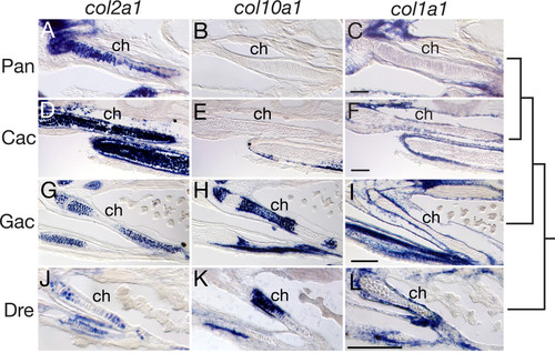
Ceratohyal collagen gene expression in P. antarcticum, C. aceratus, G. aculeatus, and D. rerio detected by in situ hybridization on cryosections. (A-C) P. antarcticum, mid-larvae; (D-F) C. aceratus, mid-larvae; (G-I) G. aculeatus, 11 dpf; (J-L) D. rerio, 6 dpf. Sections through the ceratohyal cartilage (ch) are shown in the ventral view. Notothenioid larvae show ubiquitous expression of col2a1 throughout the ch (A, D), whereas col2a1 expression is restricted to the distal ends of the cartilage in both G. aculeatus (G) and D. rerio (J). The col10a1 gene is expressed in hypertrophic chondrocytes in the ch in G. aculeatus (H) and D. rerio (K), whereas its expression is absent from the ch in P. antarcticum (B) and C. aceratus (E). The col1a1 gene is weakly expressed in the periosteum around the ch of both P. antarcticum (C) and C. aceratus (F) relative to the strong expression observed in G. aculeatus (I)and D. rerio (L). The tree represents the evolutionary relationship among species. Scale bars, 100 μm.
|
