
Fig. S6
- ID
- ZDB-FIG-100302-62
- Publication
- Seo et al., 2010 - BBS6, BBS10, and BBS12 form a complex with CCT/TRiC family chaperonins and mediate BBSome assembly
- Other Figures
- All Figure Page
- Back to All Figure Page
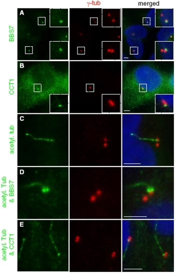
BBS7 and CCT1 specifically localize to the mature centriole. BBS7 (A) and CCT1 (B) specifically localize to only one centriole of the two in ciliated ARPE- 19 cells. The primary cilium (marked by acetylated tubulin in C) is generated from only one of the two centrioles, which is the mature or “mother” centriole and designated as the basal body. In all cases where the cilia can be unambiguously linked to one of the two centrioles, BBS7 and CCT1 staining (two green punctates in D and E) is associated with the centriole with the cilium (green whip-like structure) and within that centriole BBS7 and CCT1 are found on the side where the cilium is attached. These data indicate that BBS7 and CCT1 localize specifically to the basal body and potentially to the transition zone. However, this property does not appear to be a general feature of all BBS/CCT complex components, because we did not observe same localization in other BBS or CCT proteins tested (BBS6, BBS10, CCT4, and CCT8). |