
Fig. 3
- ID
- ZDB-FIG-100302-35
- Publication
- Loynes et al., 2010 - Pivotal Advance: Pharmacological manipulation of inflammation resolution during spontaneously resolving tissue neutrophilia in the zebrafish
- Other Figures
- All Figure Page
- Back to All Figure Page
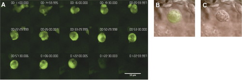
Morphological features of apoptosis are seen during time-lapse analysis of neutrophilic inflammation in zebrafish. (A) Four hours following transection of the tailfin under Tricaine anesthesia, a 4-dpf zebrafish larva was re-anesthetized and mounted in 1% low melting-point agarose containing 0.02% Tricaine. During this time-lapse series (showing a single confocal slice), a neutrophil is seen to cease movement and assume a rounded shape. This is followed by loss of GFP fluorescence. Times since initiation of time-lapse are shown (h:m:s.ms). The images have been selected to be representative of the series, which can be seen in full in Supplemental Movies 1 and 2. Photomicrographs were taken with a x40 Plan NeoFluar oil immersion objective, NA1.3 (Zeiss). As a result of the thickness of the tissue section here, the individual cells are not visible on DIC images. Exposure times were minimized, and there was no evidence of any adverse effects over 16 h of imaging. Similar events are seen under wide-field time-lapse microscopy. (B and C) A similar event occurring in the tailfin enables imaging under DIC of the persisting neutrophil cell "corpse." (B) A fluorescent neutrophil showing features of apoptotic morphology in the tailfin during inflammation resolution. (C) The same neutrophil, a single frame (30 s) later, showing that the neutrophil corpse persists, and the GFP signal has been lost completely. |