
Fig. 7
- ID
- ZDB-FIG-091216-19
- Publication
- Shi et al., 2009 - Probing events with single molecule sensitivity in zebrafish and Drosophila embryos by fluorescence correlation spectroscopy
- Other Figures
- All Figure Page
- Back to All Figure Page
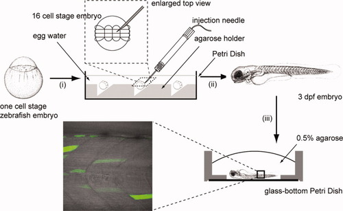
Zebrafish embryo preparation (i) one-cell stage embryos were collected and dechorionated. The embryos were aligned onto the agar holder in a Petri Dish and the orientation of the embryo was adjusted with a glass dropper that the animal pole was on top. The Petri dish was filled with egg water and the embryos were let to develop to the 16-cell stage. (ii) The DNA plasmid was microinjected into one of the four central blastomeres. The embryos were then incubated in egg water at 28.5°C to develop to 3 days postfertilization (dpf). (iii) Selected embryos were anaesthetized and mounted into 0.5% low-melting-temperature agar in a glass bottom Petri dish. The embryo body was pushed close to the surface of cover glass and the orientation of the embryo was adjusted with a needle. The specimen was then mounted in the microscope stage for subsequent confocal imaging and fluorescence correlation spectroscopy (FCS) measurements. |