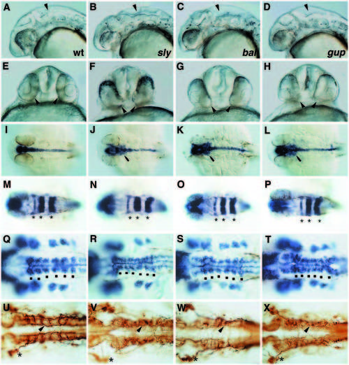
Phenotypes of mutations affecting notochord and brain. (A,E,I,M,Q,U) wild type; (B,F,J,N,R,V) sleepy (sly)m86; (C,G,K,O,S,W) bashful (bal)m190; (D,H,L,P,T,X) grumpy (gup)m189. Lateral (A,B,C,D) and anterior-ventral (E,F,G,H) view of embryos at 28 hpf. Note the enlarged hindbrain ventricles and the irregular morphology of the hindbrain (arrowhead in A-D) and the position of the eyes (arrowheads in EH). (I,J,K,L) Expression of sonic hedgehog at 29 hpf; dorsal view. Note the lateral expansion posterior to the eye in mutant embryos (arrow in J,K,L). (M,N,O,P) Expression of rtk1 in rhombomeres 1, 3 and 5 (asterisks). (Q,R,S,T) Expression orf dlx2 in pharyngeal arch primordia and hlx1 in hindbrain at 29 hpf; dorsal view. Note the slightly irregular expression domains of hlx1 adjacent to rhombomere boundaries (dots) in mutant embryos. (U,V,W,X) Medial longitudinal fascicles (arrowhead in the region of rhombomere 5), lateral longitudinal fascicles and commissures in hindbrain stained with antibody against acetylated tubulin at 29 hpf. Dorsal view. Asterisks highlight the position of the trigeminal ganglion.
|
