
Fig. 4
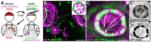
Transplanted WT central RGCs rescue retinal exit in ath5 morphants. (A) Blastula transplants from isl2b:mCherry donors resupply ath5 morphant hosts, labeled with isl2b:gfp, with early RGCs. RhDx, rhodamine-dextran cell lineage marker. (B) Resupplied WT RGCs and axons (magenta) are sufficient to rescue host axons in morphants (green). Pigment cell autofluorescence is seen around eyes and at dorsal midline (magenta). Retinal axons project across chiasm (broken lines); donor axons terminate in central tectum (asterisk), while host axons terminate in peripheral tectum (arrowheads). Dorsal view, 5 dpf. (C) Lateral view of 5 dpf eye showing peripheral host axons (green) fasciculating with resupplied WT axons (magenta). (C′) Donor and (C″) host axons, reverse contrast. Scale bars: 100 μm in B; 50 μm in C. |