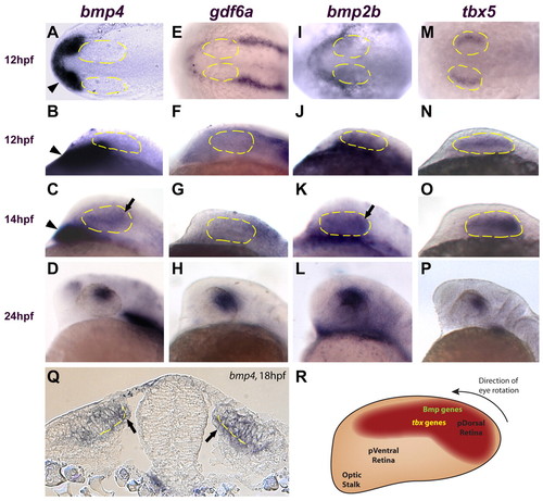
Multiple Bmp genes and tbx5 are expressed in the retina before canonical Wnt activity. (A,E,I,M) Dorsal views, anterior left. (B-D,F-H,J-L,N-P) Lateral views, dorsal up, anterior left. (A-D) bmp4 is expressed in the prechordal mesoderm at 12 and 14 hpf (arrowheads in A-C) but is not expressed in the optic vesicle until 14 hpf (arrow in C). At 24 hpf, bmp4 expression is restricted to the dorsal retina (D). (E-L) gdf6a and bmp2b are not expressed in the optic vesicle at 12 hpf (expression of these genes is restricted to the surface ectoderm). Expression of bmp2b is present in the retina at 14 hpf (arrow in K), but gdf6a does not appear in the optic vesicle until 16 hpf (not shown). Both genes are expressed in the dorsal retina at 24 hpf (H,L). (M-P) tbx5 expression begins in the optic vesicle at 12 hpf and becomes progressively restricted to the dorsal retina by 24 hpf. (Q) Transverse section through the midbrain at 18 hpf. bmp4 is expressed in the presumptive dorsal neural retina and RPE (arrows). Broken yellow lines indicate the interface between the neural retina and the RPE. (R) Diagram of the zebrafish retina at approximately 14 hpf, showing the expression domains of Bmp genes and tbx5 at this timepoint. At approximately 22 hpf, the entire eye rotates 90° in the direction indicated. Anterior left, dorsal up.
|
