
FIGURE
Fig. S3
Fig. S3
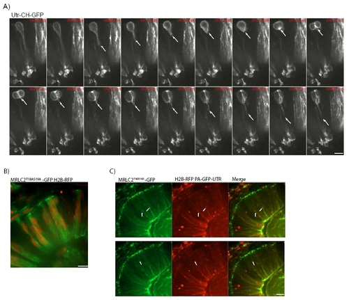
A) Images from Movie 10. GFP-Utr-CH disappears from the basal process of cells just before mitosis and cytokinesis (arrows moving up). It quickly reappears after cell division (arrows moving down). B) Non-activatable Myosin is labelled by MRLC2T18AS19A-GFP. Nuclei are labelled by H2B-RFP. C) Co-labelling of MRLC2T18DS19D-GFP for constitutively activated Myosin and RFP-Utr-CH for filamentous actin shows that localizations overlap (merge). Arrows show basal accumulation of activated myosin with actin accumulations. |
Expression Data
Expression Detail
Antibody Labeling
Phenotype Data
Phenotype Detail
Acknowledgments
This image is the copyrighted work of the attributed author or publisher, and
ZFIN has permission only to display this image to its users.
Additional permissions should be obtained from the applicable author or publisher of the image.
Reprinted from Cell, 138(6), Norden, C., Young, S., Link, B.A., and Harris, W.A., Actomyosin is the main driver of interkinetic nuclear migration in the retina, 1195-1208, Copyright (2009) with permission from Elsevier. Full text @ Cell