|
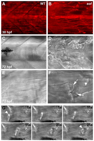
Muscle degeneration in sof mutants results from detachment of myofibres from the vertical myoseptum. Lateral views of wild-type muscle (A,C,E) show fibres spanning the somite between adjacent myosepta, whereas in sof mutants (B,D,F), lesions rapidly develop. (A,B) Whole-mount immunohistochemistry using an antibody against slow myosin demonstrates that, although slow fibres develop normally in sof embryos, by 30 hpf fibres have already detached from the myoseptum. (C-F) DIC images of axial muscle. By 72 hpf, the sof embryo appears severely damaged, with numerous detached fibres scarring the myotome. A magnified view of a single detached fibre (F) shows the characteristic invaginated membrane (arrows) and retraction groove (arrowhead). (G) Retraction of a single fibre (arrowhead), from left of frame to right, in a 72 hpf sof embryo. Still images taken from Movie 1 in the supplementary material, with elapsed time indicated in seconds.
|