
Fig. S5
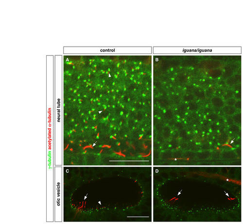
iguana mutants lack primary cilia. Cilia were visualized by staining with acetylated α-tubulin antibody (red), and basal bodies were visualized by staining with γ-tubulin antibody (green). (A,B) Primary cilia in the neural tube (arrowheads in A) are absent in igu/igu mutants (B), whereas the longer floor plate cilia (arrows) are reduced in number in igu/igu mutants as compared with control embryos (igu/+ or +/+) (A). (C,D) igu/igu mutants lack short non-motile cilia (arrowhead in C) and have a reduced number of the longer tether cilia (arrows) in the otic vesicle. Note that the cluster of tether cilia at the opposite end of the otic vesicle is out of focus in the control embryo (C). Asterisks in B and D mark the staining of axons, which also label with antibody to acetylated α-tubulin. Lateral views at 24 hpf. Scale bars: 20 μm. |