
Fig. 3
- ID
- ZDB-FIG-090717-57
- Publication
- Hultman et al., 2009 - Defects in ErbB-dependent establishment of adult melanocyte stem cells reveal independent origins for embryonic and regeneration melanocytes
- Other Figures
- All Figure Page
- Back to All Figure Page
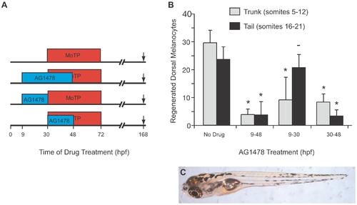
Temporal shifts of AG1478 reveal stem cell establishment occurs in a rostrocaudal progression. (A) Cartoon of drug treatment timeline. (B) Quantitation of average regenerated dorsal melanocytes for each treatment in (A) in the trunk (somites 5–12, in gray) and in the tail (somites 16–21, in black). Error bars represent standard deviation, * P<0.05, - P>0.05 (Student t-test, N = 10). Larvae not treated with AG1478 show full regeneration in the trunk and in the tail. When larvae are treated with the full AG1478 treatment, from 9–48 hpf, they fail to regenerate in either the trunk or the tail. Larvae treated early with AG1478 from 9–30 hpf fail to regenerate in the trunk, but have normal regeneration in the tail. Later treatments of AG1478 from 30–48 hpf show more regeneration in the trunk than in the tail. (C) Larva with early treatment of AG1478 from 14–24 hpf showing a regeneration defect in the trunk but with normal regeneration in the head and tail. |