
FIGURE
Fig. S4
Fig. S4
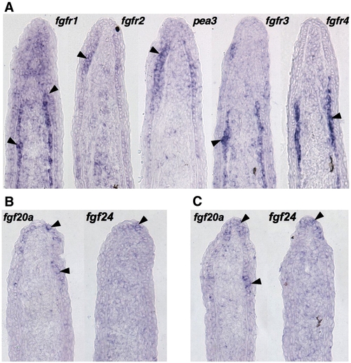
Expression patterns of fgfr and fgf genes during fin regeneration. (A) ISH of serial sections for fgfr1–4 and pea3 from a single 3 dpa (33 °C) fin regenerate, showing differential expression of receptors. fgfr1 is expressed broadly in the basal epidermis, mesenchyme, and scleroblasts, fgfr2 in the basal epidermis, and fgfr3 and fgfr4 predominantly in scleroblasts. (B, C) Two representative serial sections from 3 dpa fin regenerates (33 °C) stained by section ISH for fgf20a and fgf24. While each ligand is expressed in the basal epidermal layer, fgf20a expression extends more proximally than fgf24. |
Expression Data
Expression Detail
Antibody Labeling
Phenotype Data
Phenotype Detail
Acknowledgments
This image is the copyrighted work of the attributed author or publisher, and
ZFIN has permission only to display this image to its users.
Additional permissions should be obtained from the applicable author or publisher of the image.
Reprinted from Developmental Biology, 331(2), Lee, Y., Hami, D., De Val, S., Kagermeier-Schenk, B., Wills, A.A., Black, B.L., Weidinger, G., and Poss, K.D., Maintenance of blastemal proliferation by functionally diverse epidermis in regenerating zebrafish fins, 270-280, Copyright (2009) with permission from Elsevier. Full text @ Dev. Biol.