
Fig. 4
- ID
- ZDB-FIG-090710-6
- Publication
- Roberts et al., 2009 - Apical polarity protein PrkCi is necessary for maintenance of spinal cord precursors in zebrafish
- Other Figures
- All Figure Page
- Back to All Figure Page
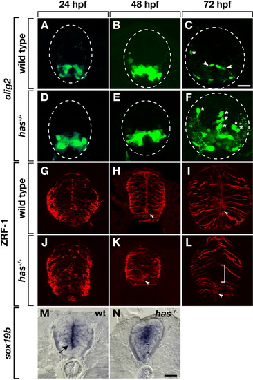
Protein kinase C, iota (PrkCi) is required to maintain ventral spinal cord cells with precursor characteristics. All images are of transverse sections through trunk spinal cord, dorsal up. Outlined circle marks the perimeter of the spinal cord. A-F: Enhanced green fluorescent protein (EGFP) expression driven by the Tg(olig2:egfp) promoter. A,B: In wild-type embryos, EGFP marks motor neurons and pMN precursors through 48 hours postfertilization (hpf). C: By 72 hpf, EGFP+ fibers (arrowheads), marking precursors that persist into larval stage, become evident. D,E: EGFP expression appears normal in has-/- embryos at 24 and 48 hpf. F: At 72 hpf, few EGFP+ radial fibers are evident. Asterisks mark dorsally migrated OPCs. G-L: Zrf-1 immunocytochemistry to label radial glial fibers. G-I: In wild-type embryos and larvae, radial fibers are distributed uniformly through the spinal cord. In ventral spinal cord, the apical membrane of some radial glia surround the central canal (arrowheads). J-L: In has mutants, radial fibers initially appear normal, but by 72 hpf, a gap appears in ventral spinal cord (bracket) and the central canal is reduced or absent. M,N: sox19b RNA expression. M: At 72 hpf in wild-type, sox19b expression marks cells near the central canal (arrow). N: At 72 hpf sox19b is not expressed at its normal position in has-/- larva (bracket). Instead, ventral and dorsal spinal cord cells express sox19b. Scale bar = 20 μM. |