|
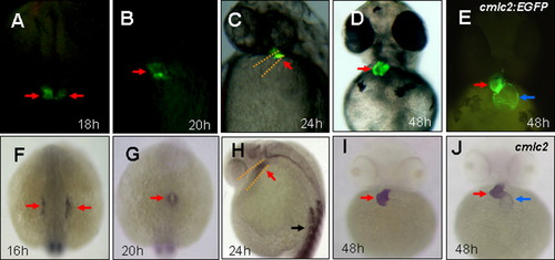
Spatial and temporal expression of vmhc:EGFP in zebrafish embryos. A-E: Fluorescent optics revealing expression of vmhc-EGFP in the lateral plate mesoderm (A), the cardiac cone (B), the ventricular portion of the heart tube (C), and the ventricle (D) in Tg[vmhc:EGFP] embryos, as well as cmlc2-EGFP expression in the ventricle and the atrium in Tg[cmlc2-EGFP] embryos (E). F-J: Whole-mount in situ analysis showing expression of vmhc in the lateral plate mesoderm (F), the cardiac cone (G), the ventricular portion of the heart tube (H), and the ventricle (I), as well as cmlc2 expression in both cardiac chambers (J). Dorsal views with anterior to the top (A,B,F,G). Lateral views (C,H). Ventral views (D,E,I,J). Red arrows, Ventricular myocytes; blue arrows, Atrial myocytes; black arrows, somites.
|