
Fig. 1
- ID
- ZDB-FIG-090618-11
- Publication
- Bouzaffour et al., 2009 - Fgf and Sdf-1 pathways interact during zebrafish fin regeneration
- Other Figures
- All Figure Page
- Back to All Figure Page
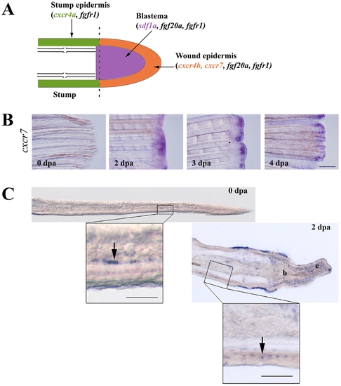
Expression of sdf1 and its receptors during fin regeneration. A: schematic representation of FGF and SDF pathways in caudal fin regeneration. In green: stump epidermis expressing cxcr4a and fgfr1. In red: wound epidermis expressing cxcr4b, cxcr7, fgf20a and fgfr1. In blue: blastema expressing sdf1a, fgf20a and fgfr1. Longitudinal cross section through the dermal ray of a regenerating fin at 2 dpa. The dotted line indicates the amputation plane. B: cxcr7 kinetics of expression during caudal fin regeneration. cxcr7 mRNA expression pattern was analyzed by in situ hybridization on control fin (0 dpa) and in amputated fins allowed to regenerate for 2, 3 and 4 dpa. Scale bar, 100 μm. C: In situ hybridization for cxcr7 on cryosections. cxcr7 mRNA expression pattern was analyzed by in situ hybridization on cryosections of uncut fin (0 dpa) and in amputated fins allowed to regenerate for 2 dpa. Two days post amputation cxcr7 mRNA is detected in the wound epidermis as well as in a few dispersed cells in the stump epidermis (enlarged view). Before amputation, only few mesenchymal cells show a staining for cxcr7. Scale bars, 50 μm. |
| Gene: | |
|---|---|
| Fish: | |
| Condition: | |
| Anatomical Terms: | |
| Stage: | Adult |