FIGURE

Fig. 1

ID
ZDB-FIG-090602-24
Publication
Warga et al., 2009 - Fate mapping embryonic blood in zebrafish: multi- and unipotential lineages are segregated at gastrulation
Other Figures
All Figure Page
Back to All Figure Page
Fig. 1

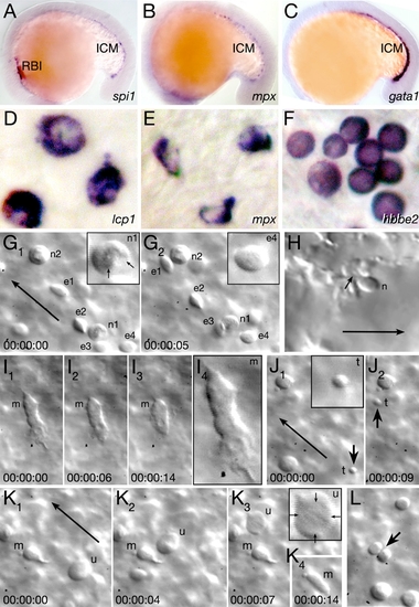
Characterization of Embryonic Blood
(A–C) The rostral blood island (RBI) and intermediate cell mass (ICM) at 18 hr, visualized with (A) a myeloid marker, (B) a neutrophil marker, and (C) an erythroid marker. (D–F) Individual blood cells at 26 hr, visualized by in situ hybridization: (D) macrophage cells, (E) neutrophil cells, and (F) erythrocyte cells. (G–L) Individual blood cells in the live 30 hr embryo. Each image is a single frame from a real-time recording of circulating blood. Elapsed time is indicated in the lower frame, long arrows indicate the flow of circulation, and insets show a 200x-magnified view of specific cells. (G and H) Neutrophils (n) and circulating erythrocytes (e) in the (G) viteline vein over the yolk sac and in the (H) lumen of a tail blood sinus. The arrows in (G1) indicate individual nuclear lobes of a neutrophil, and the small arrow in (H) indicates where the neutrophil anchors itself to the endothelial lumen. (I) A macrophage (m) patrolling the viteline vein. The last panel is magnified 200x. (J) A circulating thrombocyte (t and arrow). (K and L) Unidentified cells. (K) A large rare cell type (u), arrows in the inset indicate its diffuse nuclear envelope. A macrophage (m) is also visible in (K). (L) A dividing cell (arrow).

Expression Data
Genes:
Fish:
Anatomical Terms:
Stage Range: 14-19 somites to Prim-5

Expression Detail
Antibody Labeling
Phenotype Data

Phenotype Detail
Acknowledgments
This image is the copyrighted work of the attributed author or publisher, and ZFIN has permission only to display this image to its users. Additional permissions should be obtained from the applicable author or publisher of the image.

Reprinted from Developmental Cell, 16(5), Warga, R.M., Kane, D.A., and Ho, R.K., Fate mapping embryonic blood in zebrafish: multi- and unipotential lineages are segregated at gastrulation, 744-755, Copyright (2009) with permission from Elsevier. Full text @ Dev. Cell