
Fig. 4
- ID
- ZDB-FIG-090520-26
- Publication
- Poss et al., 2000 - Roles for Fgf signaling during zebrafish fin regeneration
- Other Figures
- All Figure Page
- Back to All Figure Page
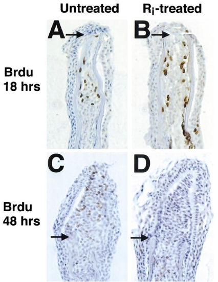
Fgfr1 inhibition blocks proliferation of established blastemal cells. (A, B) Section of 18-h fin regenerates indicating BrdU incorporation (that occurred during 12–18 h postamputation) in untreated (A) and Ri-treated (B) fins. Ri had no effects on BrdU incorporation in proximal mesenchymal tissue at this stage. Arrows demarcate amputation plane in each photograph. (C, D) Section of fins allowed to regenerate for 40 h prior to (C) a 2-h period without treatment before 6 h incubation with BrdU or (D) a 2-h Ri preincubation period before 6 h treatment with both Ri and BrdU. While proximal mesenchymal cell BrdU incorporation was normal during the brief Ri incubation, BrdU incorporation in distal blastemal cells was never observed, and incorporation in proximal blastemal cells was dramatically reduced. Original magnification was 400x. |
Reprinted from Developmental Biology, 222(2), Poss, K.D., Shen, J., Nechiporuk, A., McMahon, G., Thisse, B., Thisse, C., and Keating, M.T., Roles for Fgf signaling during zebrafish fin regeneration, 347-358, Copyright (2000) with permission from Elsevier. Full text @ Dev. Biol.